Anatomy & Physiology
|
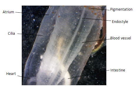
Above: Photograph of E. diaphanis zooid taken from it's ventral side, using a digital microscope.
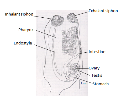
Below:Diagram of an E. diaphanis zooid from it's left side. Drawing from Kott (1985). Information adapted from Fox (2007)
|
Body Plan
Consistent with most adult ascidians, E. diaphanis zooids have a simple bilaterally symmetrical body plan with conventional axes of orientation (Harrison and Ruppert, 1991). The anterior is marked by the inhalant siphon with the neural complex on the dorsal side, close to the exhalant siphon. The inhalant siphon opens into the pharyngeal basket, which due to rows of lateral cilia becomes continuous with the atrial chamber. The endostyle extends as a long glandular groove on the ventral side of the pharyngeal basket (Harrison and Ruppert, 1991). E. diaphanis has a U-shaped gut, with a distinct esophagus, stomach, intestine and anus, with the anus terminating in the atrial chamber. The neural complex is located on the midline between the siphons, and is formed of a neural ganglion and an associated neural gland (Harrison and Ruppert, 1991). The body of an ascidian zooid is covered with a monolayered epidermis with a complex tunic enveloping the entire epidermis and lining the internal siphon surfaces (Harrison and Ruppert, 1991). The heart is positioned ventrally and posteriorly to the endostyle and the gonads are found at the anterior of the gut loop on a posterior position (Kott, 1985). A basal lamina and a thick, gelatinous connective tissue underlie the epidermis in which the muscles,nerves, blood vessels and ameboid cells are located (Harrison and Ruppert, 1991). The zooids are united by short stolons creating a clustered colony. |